The Human Eye
The eye is the organ which regulates the sense of sight, allowing us to interpret the shapes, colours and dimensions of our surroundings by processing the light they reflect or emit. This guide will take you through each step of the visual process, defining the role of the various parts and their link to each other.
Anatomy of the eye
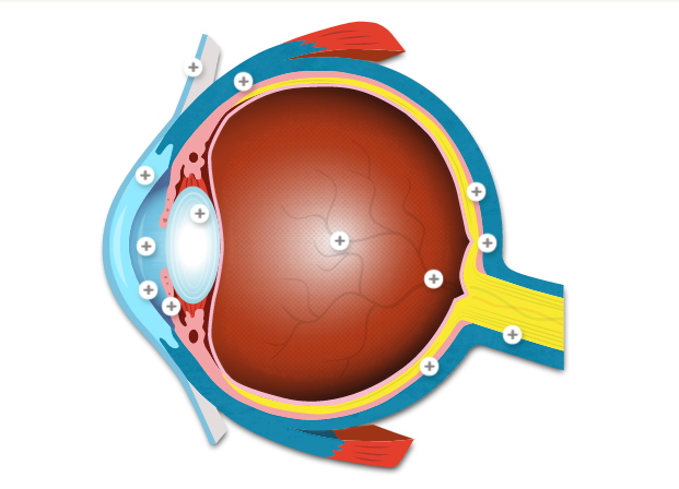
Hover over the plus buttons to learn about the functionality of each part of the eye

80% of the eyeball, a clear gel that fills the middle of the eye, and lies between the crystalline lens and the retina. Transmits the initial light waves received through the cornea, pupil and lens to the retina.
Opening in the centre of the iris that allows a certain amount of light to enter the eye. Its function is to regulate the amount of light the retina receives.
Controls the diameter and size of the pupil and thus the amount of light reaching the retina.
The transparent layer which covers the pupil, iris and anterior chamber. Its clarity is due to the fact that it is supplied with oxygen and nutrients through the tear film and not through blood vessels.
The sclera is the white, robust coat that covers the eye ball. Its function is to protect the inner, more sensitive parts of the eye. It is also connected to the 6 muscles that enable the eyeball to move and rotate.
Mucous membrane covering the sclera and the inside of the eyelids. It is responsible for part of the production of tear-fluid and prevents the entrance of microbes into the eye. When irritated, it can cause blood vessels to expand in the cornea.
Located behind the pupil, the crystalline lens focuses light rays before they reach the retina. The lens, by changing shape, functions to change the focal distance of the eye so that it can focus on objects at various distances.
Fluid filling the front part of the eye, between the lens and the cornea. Its role is to supply the cornea and the lens with oxygen and nutrients. (Originally produced at the back of the ciliary body, then seeps through the pupil, into the anterior chamber and ultimately drained through the trabecular meshwork).
Sensory membrane made of photoreceptor cells (rods and cones) which transform light impulses into electric signals
Most sensitive part of the retina, responsible for central vision
Only made of cones, the fovea is the most central part of the macula which is necessary for activities where visual detail is of primary importance, such as reading and driving.
Head of the optic nerve which is deprived of cones and rods and therefore cannot respond to light stimulation. As a result, it is also known as the blind spot. The optic disc is placed 3 to 4 mm to the nasal side of the fovea.
Connects the eye to the brain. It carries the impulses formed by the retina to the brain, which interprets them as images, transmitting all the relevant information about colours, light and darkness.
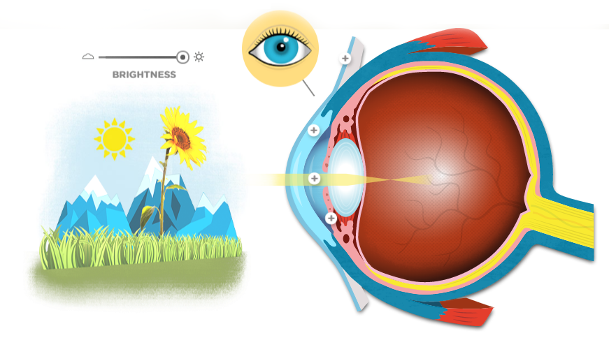
01. Light enters the eye
When you look at an external element, the light rays reflecting on this element enter the eye through the cornea which bends the light rays in such a way that

Mucous membrane covering the sclera and the inside of the eyelids. It is responsible for part of the production of tear-fluid and prevents the entrance of microbes into the eye. When irritated, it can cause blood vessels to expand in the cornea.
Transparent layer which covers the pupil, iris and anterior chamber. Its clarity is due to the fact that it is supplied with oxygen and nutrients through the tear film and not through blood vessels.
Opening in the centre of the iris that allows a certain amount of light to enter the eye. Its function is to regulate the amount of light the retina receives.
Controls the diameter and size of the pupil and thus the amount of light reaching the retina.
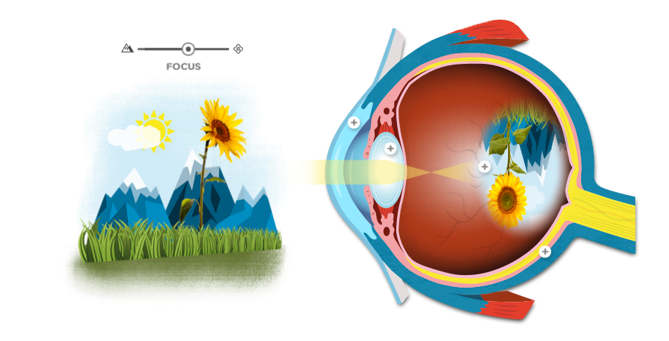
02. Lens adjusts focal length
The crystalline lens changes shape in order to adjust the focus. The resulting image appears as an upside down projection on the back of the

Transparent layer which covers the pupil, iris and anterior chamber. Its clarity is due to the fact that it is supplied with oxygen and nutrients through the tear film and not through blood vessels.
Located behind the pupil, the crystalline lens focuses light rays before they reach the retina. The lens, by changing shape, functions to change the focal distance of the eye so that it can focus on objects at various distances.
80% of the eyeball, a clear gel that fills the middle of the eye, and lies between the crystalline lens and the retina. Transmits the initial light waves received through the cornea, pupil and lens to the retina.
Sensory membrane made of photoreceptor cells (rods and cones) which transform light impulses into electric signals
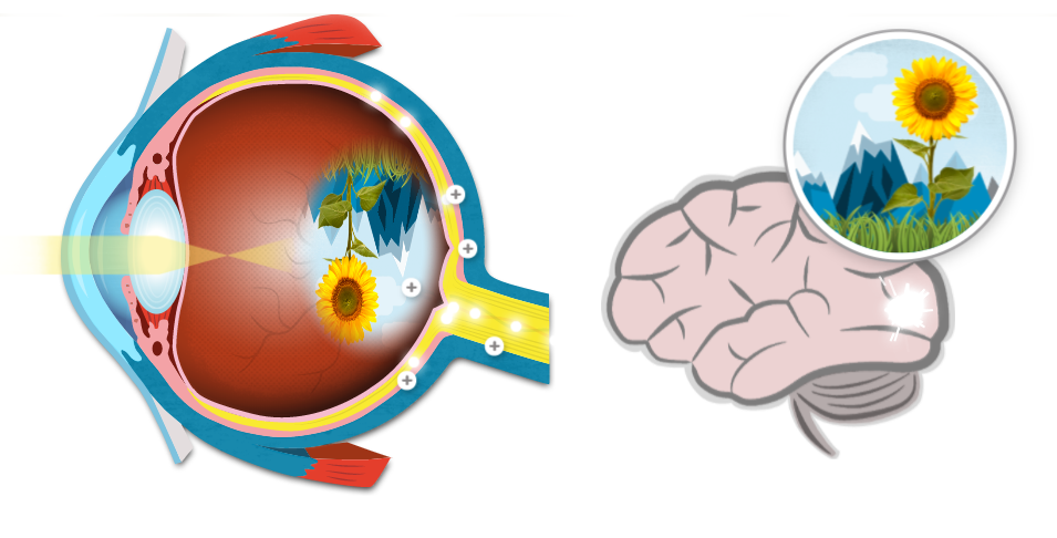
03. Brain interprets the image
When light rays reach a sharp focusing point on the retina, they are transformed into light impulses that go through a million nerve fibers to reach the optic nerve. Its role is to transmit these signals to the visual cortex which then interprets light impulses into images.

Most sensitive part of the retina, responsible for central vision
Only made of cones, the fovea is the most central part of the macula which is necessary for activities where visual detail is of primary importance, such as reading and driving.
Head of the optic nerve which is deprived of cones and rods and therefore cannot respond to light stimulation. As a result, it is also known as the blind spot. The optic disc is placed 3 to 4 mm to the nasal side of the fovea.
Connects the eye to the brain. It carries the impulses formed by the retina to the brain, which interprets them as images, transmitting all the relevant information about colours, light and darkness.
Sensory membrane made of photoreceptor cells (rods and cones) which transform light impulses into electric signals


